Use of STED microscopy to visualize liver sinusoidal endothelial fenestrae
This study was just published in Biology of the Cell by members of Team 1.
The first author of this paper is Julie di Martino, former PhD student of Fred Saltel.

- 05/06/2018
 ©F. Saltel
Biology of the cell
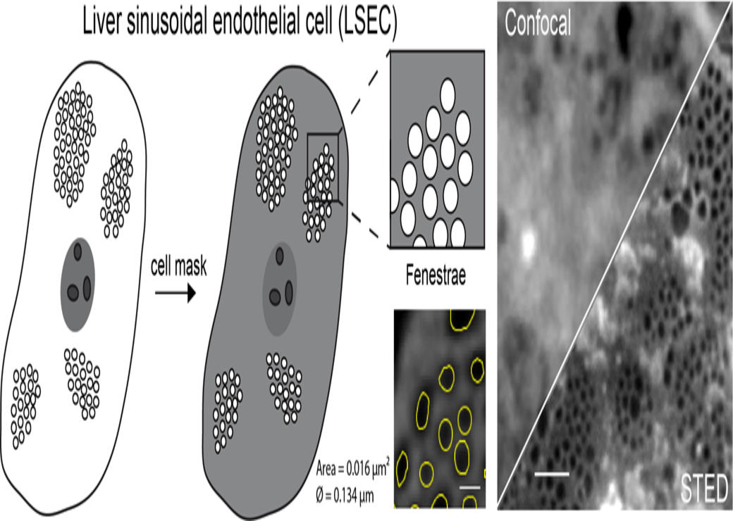
STED microscopy: a simplified method for liver sinusoidal endothelial fenestrae analysis
Julie Di Martino1,2,¶, Patrice Mascalchi2,3, Philippe Legros4, Sabrina Lacomme2,3, Etienne Gontier2,3, Paulette Bioulac-Sage1, Charles Balabaud1, Violaine Moreau1,2 and Frédéric Saltel1,2*
This study highlights that stimulated-emission-depletion (STED) microscopy is perfectly adapted for visualization, quantification, dynamics and molecular analysis of Liver sinusoidal endothelial cells (LSECs) fenestrae. Indeed, after fluorescence staining of the cell membrane or cytoplasm and using this super resolution optical microscopy method, we created a simple strategy for LSECs fenestrae analysis.